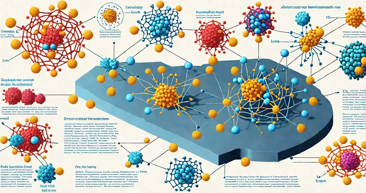
The antigen antibody reaction definition refers to the specific, high-affinity binding between an antibody and its corresponding antigen, forming an immune complex that triggers downstream immune responses. This interaction is foundational to adaptive immunity and underpins diagnostic testing, vaccine development, and autoimmune disease monitoring.
Antibodies, produced by B cells, recognize and bind to unique molecular structures called epitopes on antigens-typically proteins, polysaccharides, or lipids from pathogens like viruses or bacteria. The binding occurs through non-covalent interactions such as hydrogen bonds, van der Waals forces, and electrostatic attractions. This specificity helps antibodies do not react indiscriminately with host tissues, minimizing collateral damage.
When an antibody binds to an antigen, it can neutralize the pathogen, mark it for destruction by phagocytes, activate complement pathways, or help antibody-dependent cellular cytotoxicity (ADCC). The strength and stability of this bond depend on the affinity-the degree of attraction between a single antibody-binding site and one antigenic determinant-and avidity, which accounts for multiple simultaneous interactions in multivalent antibodies.
Each antibody has two identical antigen-binding sites, allowing it to cross-link antigens. This clustering enhances recognition by immune cells and increases the effectiveness of clearance mechanisms. For example, IgG antibodies bind to Fc receptors on macrophages, signaling the engulfment and digestion of opsonized pathogens.
Binding is reversible and concentration-dependent. At equilibrium, the formation of the antigen-antibody complex follows mass action principles. The dissociation constant (Kd) measures affinity-lower Kd values indicate tighter binding. A typical high-affinity antibody may have a Kd in the nanomolar range (e.g., 10⁻⁹ M), meaning it binds effectively even at low antigen concentrations.
In serological assays like ELISA or Western blotting, researchers exploit this reaction to detect the presence of specific antigens or antibodies. For instance, in HIV screening, a patient's serum is exposed to purified HIV envelope protein (gp120). If anti-gp120 antibodies are present, they bind and are detected using enzyme-labeled secondary antibodies, producing a visible color change.
Antigens vary in structure and origin. They include:
Antibodies fall into five major classes based on their heavy chain type:
| Type | Function | Example Use |
|---|---|---|
| IgG | Primary responder; crosses placenta; neutralizes pathogens | Most common in blood tests; detects past infections |
| IgM | First antibody produced in primary response; pentameric form | Indicates recent infection (e.g., acute hepatitis) |
| IgA | Found in mucosal secretions; prevents pathogen entry | Monitored in celiac disease via tissue transglutaminase IgA |
| IgD | Present on B cell surfaces; activates B cells | Less involved in systemic immunity |
| IgE | Binds to mast cells; triggers allergic reactions | Detects allergens in skin prick tests |
IgM antibodies have lower affinity but higher avidity due to their pentameric structure, making them effective early in infection despite weaker individual binding sites.
Many assume that all antigen-antibody reactions lead to pathogen destruction. That is incorrect. Binding alone does not eliminate threats-effector mechanisms like phagocytosis, complement activation, or ADCC are required. An antibody can bind an antigen without causing harm or benefit, especially if it targets a non-functional epitope.
Another misconception is that antibodies are always protective. In autoimmune diseases, autoantibodies mistakenly target self-antigens. For example, in type 1 diabetes, anti-insulin antibodies attack pancreatic beta cells. These reactions are harmful and illustrate that the same molecular interaction can be beneficial or destructive depending on context.
A less obvious point: antigen-antibody binding is not always irreversible. Some therapeutic monoclonal antibodies are designed to bind transiently to avoid prolonged immune activation. Rituximab, used in treating B-cell lymphomas, binds CD20 on malignant B cells and induces apoptosis through complement-mediated lysis and ADCC-but the binding itself is changing and reversible.
Consider a patient presenting with fever, rash, and joint pain. Suspected Lyme disease requires confirmation via serology. The test uses recombinant Borrelia burgdorferi outer surface protein A (OspA) as the antigen. Serum is applied to a microtiter plate coated with OspA. If anti-OspA IgM or IgG antibodies are present, they bind and are detected with enzyme-conjugated anti-human IgG/IgM.
Results are interpreted using a cutoff optical density (OD) value. A sample with OD > 0.25 is considered positive. This threshold balances sensitivity and specificity-too low increases false positives; too high misses early cases. The CDC recommends confirmatory testing with Western blot if initial results are equivocal.
This process relies entirely on the antigen antibody reaction definition: precise molecular recognition enabling accurate diagnosis. Without it, identifying infectious agents would require culturing live organisms, which takes days to weeks and fails in many cases.
Not all antigens are immunogens. An immunogen is a substance capable of inducing an immune response. Some small molecules (haptens) lack immunogenicity unless conjugated to a larger carrier protein. For example, penicillin is a hapten-it binds antibodies but cannot trigger B cell activation alone. Only when bound to serum albumin does it become immunogenic and elicit a measurable antibody response.
This explains why penicillin allergy develops only after repeated exposure: the drug must first conjugate to host proteins to form a complete antigen. The resulting immune response produces IgE antibodies against the modified protein, leading to histamine release and allergic symptoms upon re-exposure.
I recently evaluated a rapid antigen test for influenza A at a community clinic. The device uses a lateral flow strip with immobilized anti-influenza nucleoprotein antibodies. Nasopharyngeal swab fluid is applied to the sample pad. If viral antigens are present, they bind to the capture antibodies in the test zone, forming a visible line within 15 minutes.
During validation, we ran 50 samples with known PCR results. The test detected 46 true positives and missed 4 (sensitivity 92%). False positives occurred in 2 samples-likely due to cross-reactivity with similar respiratory viruses. This highlights that while the antigen antibody reaction definition provides the basis for detection, real-world performance depends on antibody quality, antigen concentration, and matrix effects in biological fluids.