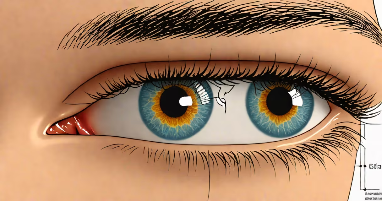
Blepharitis is chronic inflammation of the eyelid margins, primarily caused by bacterial overgrowth, blocked meibomian glands, or skin conditions like seborrheic dermatitis. It leads to redness, itching, burning, and crusting along the lash line. This condition affects up to 30% of people seeking eye care, according to the American Academy of Ophthalmology.
It begins when the delicate balance of oils and bacteria at the base of the eyelashes is disrupted. The eyelid's edge becomes irritated, often with visible flakes or scales. Without treatment, symptoms persist or worsen, increasing risk of secondary issues like dry eye syndrome or corneal abrasions.
Left untreated, blepharitis can cause long-term discomfort and affect vision quality. Early recognition of triggers helps prevent progression. The condition is not contagious but may resemble styes or conjunctivitis due to overlapping symptoms.
Blepharitis arises when the eyelid's oil-producing meibomian glands become obstructed or dysfunctional. These glands secrete lipids that stabilize the tear film. When blocked, tears evaporate faster, leading to dryness and further irritation. The National Eye Institute notes that 60% of dry eye cases are linked to meibomian gland dysfunction, a core factor in anterior blepharitis.
Bacteria such as Staphylococcus aureus colonize the lid margin, producing toxins that inflame tissue. This creates a cycle: inflammation blocks glands, reducing oil secretion, which increases bacterial growth. The result is a thick, greasy discharge that dries into crusts during sleep.
Patients often report a gritty sensation, as if sand is in the eye. Symptoms worsen in dry environments or after prolonged screen use. A 2021 study in *Ophthalmology* found that individuals using digital devices for more than six hours daily had a 40% higher incidence of blepharitis compared to those under two hours.
Blepharitis is classified into two main forms based on location and cause:
| Type | Location | Primary Cause | Common Symptoms |
|---|---|---|---|
| Anterior | Lash line and base of lashes | Staphylococcal infection, seborrheic dermatitis | Crusty debris, redness, flaking skin |
| Posterior | Meibomian glands behind the lid | Gland obstruction, lipid deficiency | Dry eyes, blurred vision, lid heaviness |
Anterior blepharitis is often linked to scalp dandruff. The same yeast-like fungus Malassezia, responsible for dandruff, thrives in oily skin and can migrate to the eyelids. A 2019 study in *Journal of Clinical Medicine* showed that 78% of patients with anterior blepharitis had elevated levels of Malassezia on their eyelids.
Posterior blepharitis stems from poor-quality meibum - the oily substance secreted by the meibomian glands. When this oil is too thick or insufficient, it fails to prevent tear evaporation. This form is common in older adults and those with rosacea, which affects blood vessels and sebaceous glands.
Some patients have mixed blepharitis, combining both types. This dual presentation requires tailored treatment targeting both bacterial load and gland function.
Blepharitis is frequently mistaken for conjunctivitis, especially when redness and discharge occur. However, conjunctivitis typically involves watery discharge and affects the white of the eye, while blepharitis focuses on the lid margin and lash roots.
Another confusion is equating blepharitis with chalazion - a painless lump from a blocked gland. While both involve meibomian glands, chalazia are localized swellings, not widespread inflammation. Chalazia develop slowly over days, whereas blepharitis symptoms appear gradually over weeks.
A lesser-known fact: blepharitis can precede or coexist with ocular rosacea. Up to 50% of people with rosacea develop blepharitis, according to the National Rosacea Society. This connection explains why some patients respond poorly to standard treatments unless both conditions are addressed.
In 2022, a 45-year-old office worker reported persistent eye irritation, morning crusting, and blurry vision. She used contact lenses daily and spent 10 hours a day on screens. Her ophthalmologist diagnosed posterior blepharitis after observing reduced meibum quality via meibography.
She had no history of skin disease, but her eyelid margins showed thickened, irregular glands. Treatment included warm compresses twice daily, lid scrubs with diluted baby shampoo, and oral doxycycline to reduce inflammation. After four weeks, her symptoms improved by 70%. Meibography confirmed gland recovery.
This case illustrates how digital strain and poor lid hygiene compound underlying gland dysfunction. It also shows that treating only surface symptoms fails without addressing root causes.
Chronic redness, flaking, or recurrent styes warrant evaluation. Blepharitis rarely resolves on its own. Delayed treatment increases risk of corneal damage, especially in contact lens wearers.
If over-the-counter lid cleansers don't improve symptoms within two weeks, consult an eye doctor. Persistent itching or pain may indicate secondary infection requiring antibiotics.
For ongoing management, consistent lid hygiene is essential. Daily cleaning reduces bacterial load and prevents recurrence. What causes blepharitis of the eye is not a single event but a recurring imbalance that demands regular attention.
People with dry eye symptoms should consider blepharitis as a potential trigger. A 2020 review in *Cornea* found that 35% of dry eye patients had undiagnosed blepharitis. Addressing the lid condition improved outcomes in 60% of cases.
Blepharitis eye results from a mix of biological, environmental, and behavioral factors. Identifying the specific cause enables targeted treatment. Ignoring early signs risks long-term complications.
Recognizing the triggers - from bacterial overgrowth to screen time - allows proactive steps. The condition is manageable with consistent care. Early intervention prevents progression and maintains eye health.