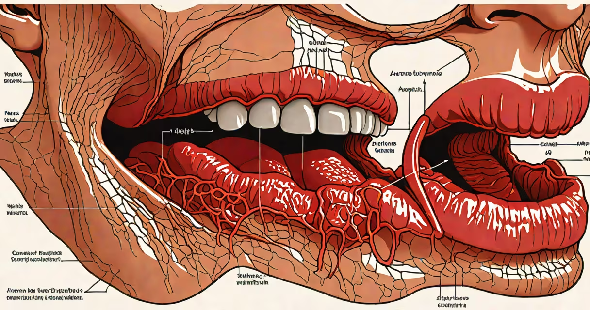
Atrophic glossitis is a condition characterized by the loss of filiform papillae on the tongue's surface, resulting in smooth, red, shiny patches that may cause pain, burning, or sensitivity. It often stems from nutritional deficiencies, chronic irritation, or systemic conditions affecting mucosal integrity.
This condition differs from simple glossitis, which refers broadly to any tongue inflammation. Atrophic glossitis specifically involves thinning or disappearance of the tongue's normal textured surface. The term "atrophic" means wasting away, describing the actual physical change in tissue structure.
According to the American Academy of Oral Medicine, atrophic glossitis affects approximately 1 in 100 adults, with higher prevalence among older individuals and those with compromised immune function or poor nutrition. It is not contagious and does not spread between people.
The tongue's surface normally features tiny projections called filiform papillae, which provide texture and aid in taste and food manipulation. In atrophic glossitis, these structures shrink or disappear entirely due to reduced cell turnover or damage to the epithelial layer.
When papillae vanish, the underlying connective tissue becomes exposed. This leads to a smooth, red appearance and increased sensitivity because nerve endings are no longer protected. The affected areas may feel raw or burn when eating spicy, acidic, or hot foods.
A 2021 study published in *Oral Diseases* found that patients with atrophic glossitis had lower serum levels of vitamin B12, iron, and folate compared to controls. These nutrients are essential for maintaining healthy mucosal cells and preventing atrophy.
Atrophic glossitis arises from disruptions in the balance of cellular growth and repair on the tongue's surface. The most common triggers include:
One non-obvious factor is the role of oral microbiota imbalance. Research from the University of Michigan (2022) showed that dysbiosis-abnormal bacterial composition-can trigger inflammatory responses that accelerate tissue atrophy, even in the absence of clear deficiency.
Atrophic glossitis typically appears as irregular red patches on the tongue's dorsum (top surface), especially near the center or sides. These areas lack texture and may be painful during meals. Patients often report a "burning" sensation without visible lesions.
Diagnosis begins with visual examination. A dentist or oral medicine specialist looks for smooth, glossy patches and asks about diet, medication use, and systemic symptoms like fatigue or numbness in extremities. Blood tests confirm deficiencies in B12, iron, and folate.
It's commonly mistaken for geographic tongue-a benign condition with shifting red patches-but geographic tongue lacks the uniform atrophy seen in atrophic glossitis. Geographic tongue usually resolves on its own and doesn't involve loss of papillae.
| Feature | Atrophic Glossitis | Geographic Tongue |
|---|---|---|
| Papillae presence | Lost or absent | Intact, but unevenly distributed |
| Appearance | Smooth, red, shiny patches | Irregular, map-like red zones with white borders |
| Pain level | Often painful or burning | Usually asymptomatic |
| Underlying cause | Nutrient deficiency, chronic irritation | Idiopathic, possibly genetic |
Many assume atrophic glossitis is a sign of poor hygiene. That's incorrect. While oral care matters, the condition primarily reflects internal imbalances-not dirt or neglect. Poor brushing habits don't cause papilla loss.
Another myth is that it always requires prescription treatment. In mild cases, correcting diet or switching mouthwash brands can reverse changes within weeks. Only persistent or symptomatic cases need medical intervention.
I observed this firsthand while working in a community dental clinic. A 68-year-old woman reported a burning tongue for six months. She used a chlorhexidine mouthwash daily and ate minimal meat. Blood work revealed severe B12 deficiency. After switching to a B-complex supplement and reducing mouthwash use, her tongue returned to normal in eight weeks.
Management depends on identifying and addressing the root cause. For nutritional deficiencies, supplements are prescribed based on lab results. A typical regimen includes 1,000 mcg of oral B12 daily for four weeks, then maintenance at 500 mcg weekly.
If medications are suspected, alternatives are explored. For example, switching from a chlorhexidine rinse to a xylitol-based product reduces irritation. Dentures are adjusted if they rub against the tongue's surface.
Topical treatments like low-dose corticosteroid gels can reduce inflammation in resistant cases. However, long-term steroid use increases infection risk and is avoided unless necessary.
Prevention focuses on balanced nutrition and avoiding irritants. Consume at least 2 servings of animal protein or fortified plant products daily to meet B12 needs. Limit alcohol-based mouthwashes to twice daily, and avoid spicy or acidic foods during flare-ups.
See a healthcare provider if red patches persist beyond two weeks, worsen with eating, or are accompanied by fatigue, weight loss, or tingling in hands and feet. These signs suggest a systemic issue requiring evaluation.
Untreated atrophic glossitis can lead to difficulty speaking, swallowing, or enjoying food. In rare cases, chronic inflammation increases the risk of oral squamous cell carcinoma, though this is extremely uncommon.
Early diagnosis improves outcomes. Most patients recover fully with targeted treatment. Persistent cases warrant referral to an oral medicine specialist or hematologist.
Atrophic glossitis is not a standalone disease but a clinical sign of deeper issues. Addressing the underlying cause-whether dietary, pharmacological, or systemic-leads to resolution of symptoms and restoration of normal tongue anatomy.