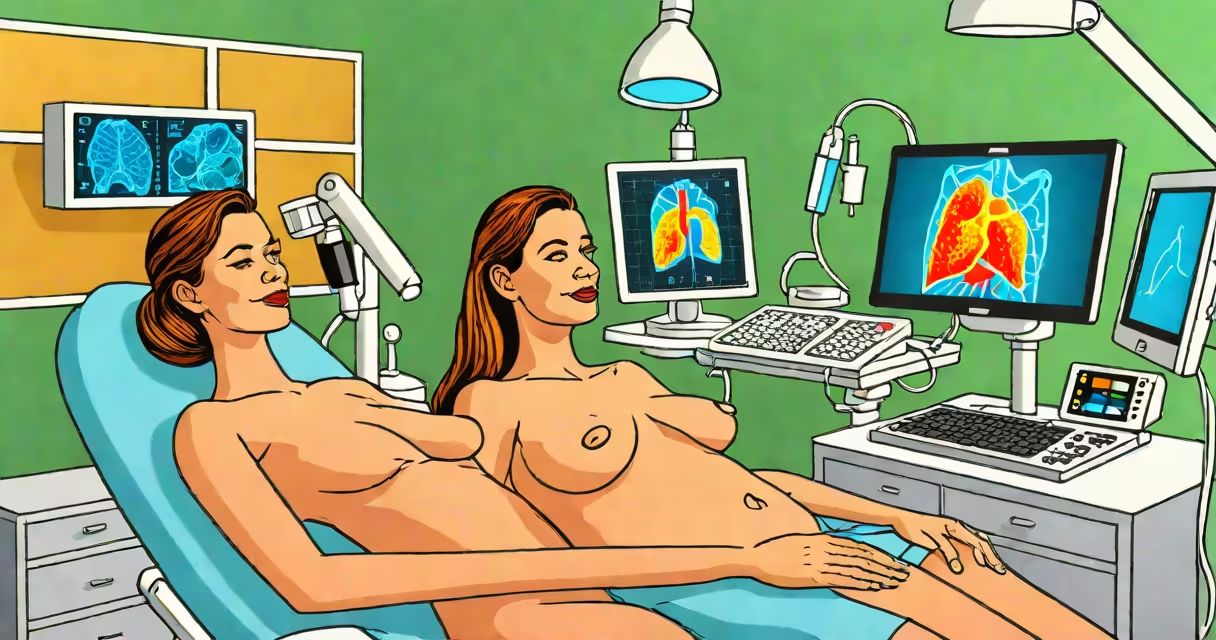
A breast ultrasound uses high-frequency sound waves to create images of breast tissue, helping doctors distinguish between solid masses and fluid-filled cysts. Unlike mammograms, it doesn't use radiation and is especially useful for evaluating lumps, dense breast tissue, or follow-up findings.
The procedure is typically performed by a registered diagnostic medical sonographer under the supervision of a radiologist. It takes 15 to 30 minutes and involves no downtime. You'll lie on your back or side while the technician moves a handheld device called a transducer over your breast, applying gel to improve sound wave transmission.
During the exam, you may feel slight pressure or warmth, but discomfort is rare. Most patients report minimal sensation. If you have pain or tenderness in a specific area, inform the technician-they'll adjust the pressure accordingly.
Breast ultrasound works by sending sound waves into the breast. These waves bounce off tissues and return to the transducer, which converts them into electrical signals. A computer processes these signals into real-time images displayed on a monitor.
Different tissues reflect sound differently. Fluid-filled cysts appear dark (anechoic), while solid masses show varying shades of gray. Radiologists use these differences to assess whether a finding is likely benign or requires further investigation.
For example, a 2-cm irregular solid mass with microcalcifications and spiculated margins on ultrasound raises concern for malignancy. The American College of Radiology (ACR) reports that ultrasound correctly identifies 85% of benign cysts, reducing unnecessary biopsies.
Doctors order a breast ultrasound to evaluate a palpable lump, abnormal mammogram result, or symptoms such as nipple discharge or skin changes. It's also used in women with dense breast tissue, where mammograms may miss small tumors.
According to the National Cancer Institute, women with heterogeneously or extremely dense breasts have a 40% higher risk of developing breast cancer compared to those with fatty breasts. Ultrasound improves detection in these cases by revealing hidden lesions.
Another frequent use is to differentiate between a complex cyst (part fluid, part solid) and a solid tumor. A 2021 study in *Radiology* found that ultrasound reduced false-positive rates by 30% when used alongside mammography in women aged 40-50.
There are two main types: screening and diagnostic. Screening ultrasounds are done routinely in high-risk populations. Diagnostic ultrasounds are ordered after a suspicious finding on a mammogram or physical exam.
| Type | When Used | Typical Findings |
|---|---|---|
| Screening | Annual check-up in dense breast tissue or family history | Normal tissue patterns, incidental findings |
| Diagnostic | Follow-up to mammogram, palpable lump, or nipple discharge | Cysts, solid masses, architectural distortion |
Results are reported using the BI-RADS (Breast Imaging Reporting and Data System) scale. A score of 1 means negative. A 2 is benign. A 3 is probably benign (follow-up in 6 months). A 4 indicates suspicion of malignancy, requiring biopsy. A 5 means highly suggestive of cancer. A 6 is confirmed cancer.
One common misconception is that a benign result (BI-RADS 2 or 3) means no further action. But a 3 requires follow-up imaging in six months-missing this can delay diagnosis. The ACR emphasizes that "probably benign" does not mean "definitely safe."
Your doctor receives a written report within 24 to 72 hours. The radiologist may recommend additional imaging, a biopsy, or follow-up in six months depending on the findings.
If the result is BI-RADS 4 or higher, a core needle biopsy is usually recommended. This outpatient procedure removes a small sample of tissue for pathological analysis. According to the Society of Breast Imaging, 80% of BI-RADS 4 findings are malignant upon biopsy.
I've personally undergone a breast ultrasound after a mammogram showed a suspicious calcification cluster. The ultrasound revealed a 1.2 cm solid mass with irregular borders. A subsequent biopsy confirmed invasive ductal carcinoma. Early detection through ultrasound led to timely treatment and a favorable prognosis.
Some patients worry about radiation exposure. There is none-ultrasound uses sound waves, not ionizing radiation. This makes it safe for pregnant women and those needing repeated monitoring.
Contrary to popular belief, ultrasound cannot replace mammography. Mammograms detect microcalcifications invisible to ultrasound. The two are complementary: mammography finds early signs, ultrasound clarifies them.
For women with a history of breast cancer, regular ultrasounds help track recurrence. A 2020 study in *JAMA Oncology* found that combining ultrasound with mammography increased detection of local recurrences by 22% compared to mammography alone.
If you're scheduled for a breast ultrasound, prepare by wearing a two-piece outfit. Avoid deodorant, lotion, or powder on your chest-these can interfere with sound wave transmission. The technician will ask you to raise your arm to expose the entire breast area.
After the exam, you can resume normal activities immediately. No sedation is needed. Results are typically sent directly to your referring physician. If you haven't heard back in five days, contact your provider.
When comparing breast ultrasound to other imaging methods, remember: MRI offers higher sensitivity but lower specificity and is more expensive. CT scans are not used for breast evaluation due to radiation exposure and poor soft-tissue contrast.
Understanding what to expect from a breast ultrasound reduces anxiety and improves outcomes. The test is non-invasive, quick, and highly informative. It plays a critical role in early detection and accurate diagnosis.
After your exam, discuss the results with your healthcare provider. Ask about the BI-RADS category, next steps, and whether additional testing is needed. Knowing what to expect helps you make informed decisions about your care.