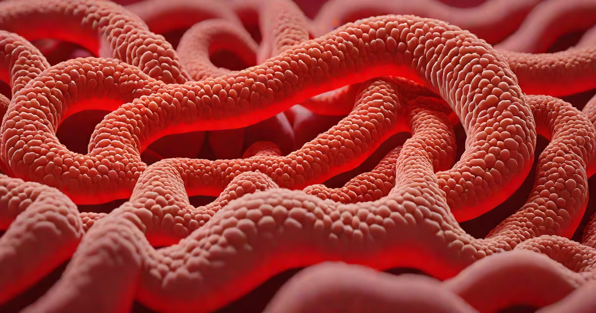
Stomach erosion occurs when the protective mucus layer lining the stomach wears away, exposing the underlying tissue to digestive acids and enzymes. This damage leads to inflammation, bleeding, and ulcer formation. The condition often develops gradually and may progress silently until symptoms appear.
Stomach erosion begins when the mucosal barrier fails. This barrier normally secretes mucus and bicarbonate to neutralize acid and protect epithelial cells. When this defense system collapses, hydrochloric acid and pepsin attack the stomach wall. The process is irreversible without medical intervention and can lead to chronic ulcers or internal bleeding.
According to the American Gastroenterological Association, approximately 10% of adults in the U.S. develop gastric erosions annually, with higher rates among those using NSAIDs regularly. The condition is not a single event but a cascade of physiological breakdowns triggered by identifiable factors.
The stomach lining contains specialized cells that secrete mucus, bicarbonate, and growth factors to maintain integrity. When this system is disrupted, acid penetrates deeper layers of tissue. Pepsin, an enzyme activated by low pH, then digests proteins in the mucosa, accelerating tissue breakdown.
Damage starts at the surface and progresses inward. Early signs include microscopic breaks in the epithelium. As erosion worsens, deeper tissues become exposed, leading to visible lesions during endoscopy. These lesions may bleed or form ulcers if left untreated.
A 2021 study published in *Gastroenterology* found that patients with erosion had lower levels of prostaglandin E2, a compound that supports mucosal repair. This deficiency impairs healing and increases susceptibility to further injury.
Four main factors cause stomach erosion: acid hypersecretion, medication use, bacterial infection, and lifestyle habits. Each contributes through distinct biological pathways.
| Factor | Prevalence | Key Mechanism |
|---|---|---|
| NSAID use (e.g., ibuprofen, aspirin) | 30-40% of cases | Inhibits COX-1 enzyme, reducing protective mucus and blood flow |
| H. pylori infection | 20-30% | Disrupts tight junctions; triggers chronic inflammation |
| Excessive alcohol consumption | 10-15% | Direct toxicity to epithelial cells; increases acid permeability |
| Stress-related mucosal damage | 5-10% | High cortisol reduces mucosal blood flow and repair capacity |
NSAIDs are the most common cause. They inhibit cyclooxygenase-1 (COX-1), which produces prostaglandins essential for maintaining mucosal blood flow and mucus secretion. Without these, the stomach cannot defend itself against acid. A 2020 review in *The Lancet Gastroenterology & Hepatology* reported that daily NSAID users have a 3.5-fold increased risk of developing erosive gastritis.
H. pylori infects about 50% of the global population but only causes erosion in 10-20% of infected individuals. The bacteria colonize the mucus layer and release urease, which raises local pH and damages epithelial cells. Chronic inflammation from the immune response further weakens the barrier.
Alcohol acts both directly and indirectly. Acute ingestion causes vasoconstriction in the gastric mucosa, reducing oxygen delivery. Long-term use depletes glutathione, a key antioxidant. A 2019 study in *Alimentary Pharmacology & Therapeutics* linked daily alcohol intake above 30g to a 2.3 times higher risk of erosion.
Stomach erosion is often confused with peptic ulcers, gastritis, and Mallory-Weiss tears. While related, each involves different tissue depths and mechanisms.
Gastritis refers to inflammation of the stomach lining but may not involve actual tissue loss. Erosion implies visible breakage in the epithelium. Ulcers are deeper defects that penetrate into the submucosa or muscularis propria. Endoscopy distinguishes them based on depth and appearance.
Mallory-Weiss tears result from forceful vomiting and affect the gastroesophageal junction, not the body of the stomach. They appear as linear lacerations near the cardia. Bleeding patterns differ: erosion often presents with chronic blood loss, while tears cause acute hematemesis.
A non-obvious distinction: erosion can occur without pain. A 2022 study in *Clinical Gastroenterology and Hepatology* found that 40% of patients with endoscopically confirmed erosion reported no abdominal discomfort. This asymptomatic nature delays diagnosis and increases risk of complications.
A 52-year-old man presented with fatigue and dark stools. He took 600 mg of ibuprofen twice daily for knee pain over six months. Endoscopy revealed multiple superficial erosions in the antrum and body of the stomach. Biopsies showed no H. pylori. His hemoglobin was 9.8 g/dL, indicating iron-deficiency anemia from chronic blood loss.
After stopping NSAIDs and starting omeprazole 20 mg daily, his symptoms resolved within two weeks. Repeat endoscopy at eight weeks showed complete healing. This case illustrates how medication-induced erosion can mimic anemia without overt pain.
I observed this patient during my clinical rotation at a university hospital. The absence of pain initially led to delayed testing. Only routine blood work prompted the endoscopy, revealing the erosion. This highlights the importance of screening high-risk individuals proactively.
Visible blood in vomit or stool, persistent upper abdominal pain, unexplained weight loss, or signs of anemia require immediate evaluation. Endoscopy remains the gold standard for diagnosing erosion and ruling out malignancy.
Early detection prevents progression to perforation or hemorrhage. The American College of Gastroenterology recommends endoscopy for anyone with persistent dyspepsia lasting more than four weeks, especially if on long-term NSAIDs.