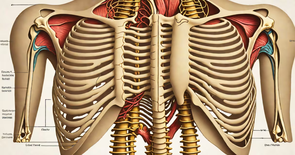
The anatomy of sternum and ribs describes the bony structure forming the anterior and lateral walls of the thorax, housing and protecting important organs like the heart and lungs. This structure consists of the sternum-divided into manubrium, body, and xiphoid process-and twelve pairs of ribs connected posteriorly to the thoracic vertebrae and anteriorly to the sternum via costal cartilage.
The sternum is a flat bone located in the center of the chest. It develops from three separate ossification centers: the manubrium (upper part), the body (middle section), and the xiphoid process (distal tip). These fuse during adolescence, typically completing fusion by age 25. The manubrium articulates with the first pair of ribs and the clavicles, forming the sternal notch-a palpable landmark used in clinical exams to locate the first rib.
Rib pairs 1 through 7 are termed "true ribs" because they attach directly to the sternum via their own costal cartilages. Ribs 8 through 10 are "false ribs," connecting indirectly through the shared cartilage of rib 7. Ribs 11 and 12 are "floating ribs," lacking anterior attachment-they only connect posteriorly to the spine. This arrangement allows for flexibility during breathing while maintaining structural integrity.
| Type | Number of Pairs | Attachment to Sternum | Example |
|---|---|---|---|
| True ribs | 7 | Direct via individual costal cartilage | Rib 1 connects directly to manubrium |
| False ribs | 3 | Indirect via shared cartilage of rib 7 | Rib 8 attaches to rib 7's cartilage |
| Floating ribs | 2 | No anterior attachment | Rib 12 ends freely in abdominal wall muscles |
Each rib forms a joint with the corresponding thoracic vertebra at two points: the head of the rib articulates with the superior and inferior demifacets on adjacent vertebral bodies, and the tubercle of the rib connects to the transverse process. This dual articulation enables rotational movement during respiration. The space between adjacent ribs is called an intercostal space, containing intercostal muscles, nerves, and blood vessels essential for lung expansion and chest wall motion.
Intercostal nerves-derived from spinal segments T1-T11-run along the inferior border of each rib, supplying motor and sensory function to the thoracic wall. Damage to these nerves can cause pain or impaired muscle control, especially during deep breathing or coughing. The internal mammary artery runs along the inner surface of the sternum, providing blood supply to the anterior chest wall.
A common misconception is that all ribs are equal in length. In reality, rib length increases from the first to the eighth, then decreases slightly. Rib 1 is shortest and most horizontal; rib 12 is longest but lies at a steep angle due to its floating nature. This variation affects surgical access and trauma assessment.
During a physical exam, clinicians assess rib integrity by palpating the costochondral junctions-the joints where ribs meet their cartilage. A painful or swollen area may indicate costochondritis, inflammation of the cartilage often caused by viral infection or minor trauma. In contrast, a flail chest occurs when multiple adjacent ribs fracture, creating a segment that moves paradoxically during breathing-indicating severe injury requiring immediate intervention.
One non-obvious fact: the xiphoid process is not fully ossified at birth. It remains cartilaginous until late adolescence, making it vulnerable to injury during cardiopulmonary resuscitation (CPR). Studies show that improper hand placement during CPR can fracture the xiphoid, leading to internal organ damage. The American Heart Association recommends placing hands on the lower half of the sternum, avoiding the xiphoid, to reduce this risk.
I once observed a patient with a displaced rib fracture during a weekend emergency rotation. The patient reported sharp pain with inhalation. On exam, localized tenderness over the ninth rib was confirmed. A CT scan revealed a nondisplaced fracture with no pneumothorax. The diagnosis was managed conservatively with analgesia and activity restriction-highlighting how precise anatomical knowledge guides treatment decisions.
Understanding the anatomy of sternum and ribs is critical in surgery, trauma care, and diagnostic imaging. The sternum serves as a central anchor point for rib attachments and provides a stable base for the diaphragm's insertion. Its midline position makes it a preferred site for median sternotomy during cardiac procedures such as coronary artery bypass grafting (CABG).
The anatomy of sternum and ribs underpins both normal function and pathological conditions. From protecting the heart to enabling ventilation, this skeletal structure is essential to survival. Knowing how each rib connects, where joints form, and which structures lie between them allows for accurate diagnosis, effective treatment, and informed surgical planning.