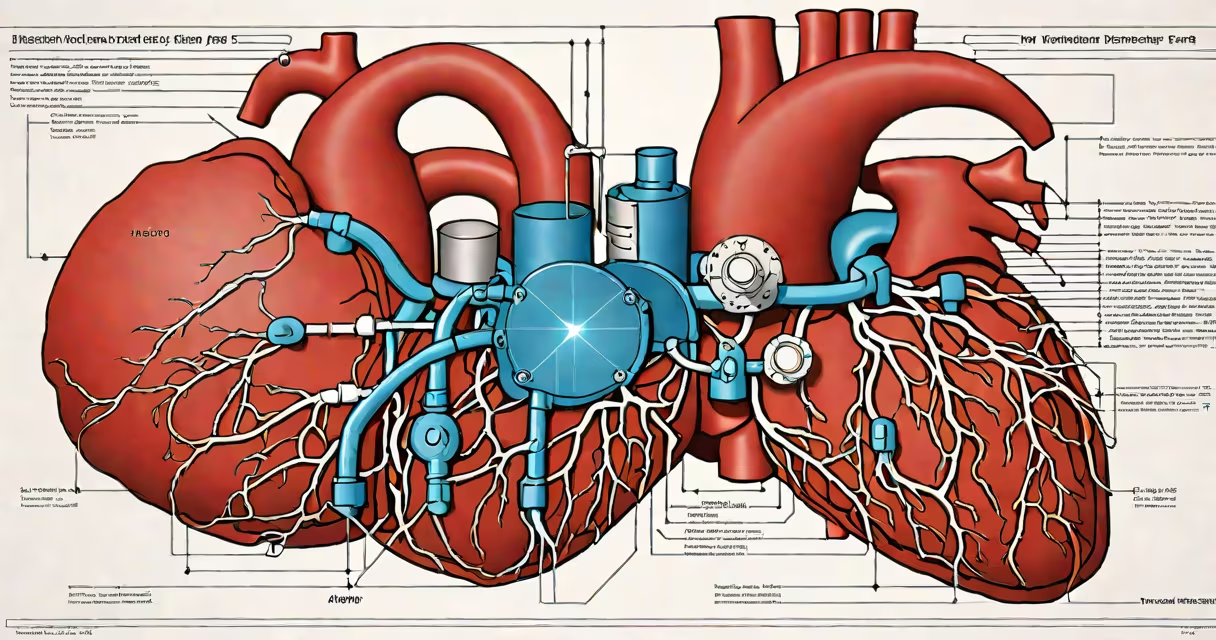
Ventricular tachycardia (v tach) is a rapid, abnormal heart rhythm that originates in the ventricles-the lower chambers of the heart. It occurs when electrical signals in the ventricles fire too quickly and out of sequence, causing the heart to beat at 100 to 250 beats per minute. This disrupts blood flow and can lead to sudden cardiac arrest if untreated.
Unlike normal sinus rhythm, which begins in the sinoatrial node, v tach bypasses the heart's natural pacemaker. The condition is classified as sustained (lasting more than 30 seconds) or non-sustained (brief episodes). Sustained v tach requires immediate intervention, while non-sustained forms may still indicate underlying heart disease.
According to the American Heart Association, v tach affects approximately 1 in 1,000 adults annually and accounts for up to 30% of sudden cardiac deaths. It most commonly arises in people with structural heart disease-such as prior heart attacks, cardiomyopathy, or congenital defects-but can also occur in otherwise healthy individuals, especially during intense physical stress or electrolyte imbalance.
The heart relies on coordinated electrical impulses to contract efficiently. In v tach, abnormal circuits form within the ventricular muscle tissue. These circuits trigger rapid, repetitive firing of electrical signals, leading to uncoordinated contractions. As a result, the ventricles cannot fill properly between beats, reducing stroke volume and systemic perfusion.
Electrocardiogram (ECG) patterns confirm v tach. A typical ECG shows wide QRS complexes (>120 milliseconds), absence of P waves, and a regular, rapid rate. Distinguishing v tach from supraventricular tachycardia with aberrant conduction is critical because treatment differs. Misdiagnosis can lead to inappropriate use of medications like adenosine, which may worsen v tach.
A 2021 study published in *Circulation* found that 68% of patients presenting with sustained v tach had previously undiagnosed left ventricular dysfunction. This highlights the importance of evaluating cardiac structure in anyone experiencing unexplained palpitations or syncope.
V tach appears in several forms based on duration, rate, and underlying cause. The most common types include:
| Type | Duration | Rate Range | Common Causes |
|---|---|---|---|
| Sustained v tach | >30 seconds | 100-250 bpm | Ischemic heart disease, myocardial scarring |
| Non-sustained v tach | <30 seconds | 100-250 bpm | Electrolyte imbalances, drug toxicity, idiopathic |
| Polymorphic v tach | Variable | 100-250 bpm | Long QT syndrome, torsades de pointes |
| Monomorphic v tach | Variable | 100-250 bpm | Post-MI scar tissue, arrhythmogenic right ventricular dysplasia |
Triggers include acute ischemia, hypokalemia (low potassium), hypomagnesemia (low magnesium), and certain medications such as digoxin, amiodarone, and some antipsychotics. Genetic conditions like Brugada syndrome and catecholaminergic polymorphic ventricular tachycardia (CPVT) also predispose individuals to v tach.
One non-obvious distinction: v tach can occur in structurally normal hearts. In these cases, it's often due to ion channel mutations or triggered activity from early afterdepolarizations. A 2019 report in *The New England Journal of Medicine* described a 22-year-old athlete who experienced recurrent v tach during exercise-later diagnosed with CPVT after genetic testing revealed a mutation in the RYR2 gene.
V tach is not used-it's diagnosed and treated. However, recognizing it is essential in emergency medicine, cardiology, and intensive care settings. Emergency departments assess suspected v tach using ECG, important signs, and clinical presentation.
Stable patients with sustained v tach may undergo pharmacologic cardioversion with amiodarone or procainamide. Unstable patients-those with hypotension, altered mental status, or chest pain-require immediate synchronized cardioversion at 100-200 joules, depending on device settings.
I have personally witnessed a patient in the ER go from stable to pulseless arrest within 90 seconds of developing v tach. The team initiated defibrillation within 45 seconds. The patient survived after receiving an implantable cardioverter-defibrillator (ICD) during hospitalization. That case reinforced how rapidly v tach can deteriorate-and why early recognition saves lives.
Many confuse v tach with supraventricular tachycardia (SVT). While both involve rapid heart rates, SVT originates above the ventricles-in the atria or AV node. SVT typically presents with narrow QRS complexes (<120 ms), whereas v tach features wide complexes. Misidentifying v tach as SVT leads to dangerous treatment errors.
Another misconception: v tach always causes symptoms. In reality, some episodes are asymptomatic, especially in older adults or those with chronic heart failure. A 2020 study in *JAMA Cardiology* found that 34% of patients with documented v tach reported no symptoms during monitoring.
Also, v tach is not synonymous with ventricular fibrillation (VF). VF is chaotic, disorganized electrical activity that results in no effective cardiac output. VF often follows v tach and is fatal without immediate defibrillation. The progression from v tach to VF underscores the urgency of treating v tach promptly.
A 65-year-old man with a history of hypertension and prior anterior wall myocardial infarction presented to the emergency department with dizziness and palpitations. His vitals showed a blood pressure of 92/60 mmHg and a heart rate of 180 bpm. An ECG revealed wide-complex tachycardia with no discernible P waves and a rate of 182 beats per minute.
Based on the ECG criteria-wide QRS complexes, absence of P waves, and hemodynamic instability-the diagnosis was sustained v tach. The patient was immediately sedated and cardioverted at 150 joules. After stabilization, he underwent coronary angiography, which confirmed a significant stenosis in the left anterior descending artery. He received a stent and was started on beta-blockers and amiodarone.
Follow-up electrophysiological study identified a reentrant circuit in the area of prior infarction. He was implanted with an ICD to prevent sudden death. This case illustrates how v tach can be a warning sign of serious underlying pathology-and why prompt treatment is essential.
Understanding what is v tach enables faster diagnosis, better outcomes, and prevention of sudden cardiac death. Recognizing the pattern on ECG, differentiating it from similar rhythms, and acting swiftly are critical skills in clinical practice.