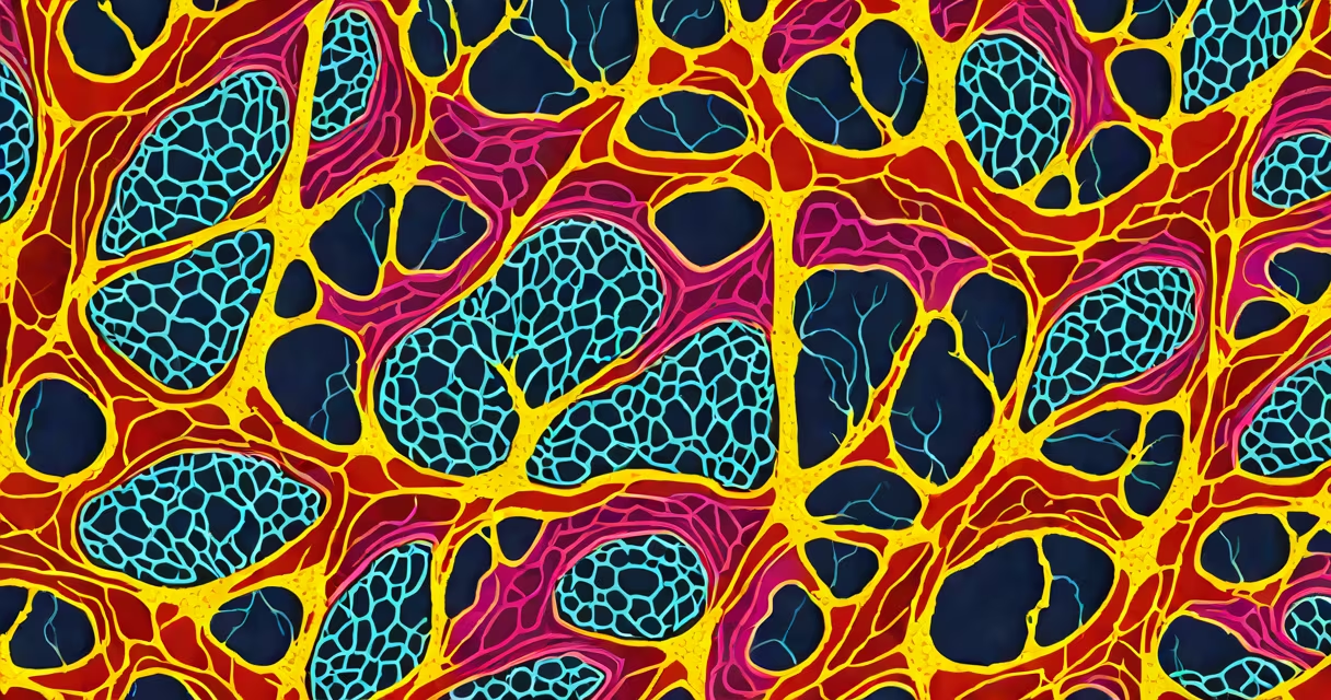
Connective tissue is defined by its abundant extracellular matrix, diverse cell types, and origin from mesenchyme. It supports, binds, and protects organs while enabling nutrient exchange and structural resilience across the body.
Connective tissue forms the structure of the body. Unlike epithelial or muscle tissues, it contains minimal cells relative to the extracellular matrix. This matrix consists of fibers-primarily collagen and elastin-and a gel-like ground substance. Fibroblasts, the most common cell type, synthesize and maintain this matrix. The tissue's structure enables functions ranging from shock absorption in cartilage to mechanical strength in tendons.
Connective tissue arises from mesenchymal stem cells during embryonic development. These undifferentiated cells migrate throughout the embryo and differentiate into fibroblasts, chondrocytes, osteocytes, and adipocytes. This shared origin explains the functional diversity within the category-from blood (a fluid connective tissue) to bone (a rigid form).
The defining features of connective tissue are its matrix composition, cellular diversity, and vascularization. The matrix is not passive-it actively regulates cell behavior through signaling molecules embedded in the ground substance. Collagen fibers provide tensile strength; elastin allows stretch and recoil. Glycosaminoglycans (GAGs) in the ground substance bind water, creating a hydrated gel that resists compression.
Cellularity varies widely. Loose connective tissue like areolar tissue contains many fibroblasts, macrophages, and mast cells. Dense regular connective tissue in tendons has fewer cells but highly aligned collagen fibers. Adipose tissue is dominated by lipid-storing adipocytes, with sparse stromal cells. This variation reflects functional specialization: loose tissue supports immune surveillance, dense tissue transmits force, and adipose tissue stores energy.
Most connective tissues are well-vascularized, allowing rapid delivery of oxygen and nutrients. Blood is the exception-its matrix is liquid plasma, and it lacks a solid fiber network. Yet it still qualifies as connective tissue due to its mesenchymal origin and role in transport and defense.
Connective tissues fall into four broad categories based on matrix density and fiber type:
| Type | Matrix Density | Fiber Types | Primary Function | Example Location |
|---|---|---|---|---|
| Loose connective tissue | Low | Collagen, elastic | Support, immune response | Dermis, around blood vessels |
| Dense regular | High | Collagen only | Force transmission | Tendons, ligaments |
| Dense irregular | High | Collagen only | Resistance to multidirectional stress | Deep dermis, organ capsules |
| Adipose | Low | Minimal | Energy storage, insulation | Subcutaneous layer, around kidneys |
| Cartilage | Moderate | Collagen, elastic | Structural support with flexibility | Joint surfaces, nose, trachea |
| Bone | Very high | Collagen, calcium salts | Load-bearing, mineral storage | Skull, femur, vertebrae |
| Blood | Fluid | None | Transport, immunity | Arteries, veins, capillaries |
Each type serves distinct roles. For example, the calcified matrix of bone provides rigidity, while the proteoglycan-rich matrix of hyaline cartilage resists compressive forces in joints. The presence of specific proteins determines function: type II collagen dominates in cartilage, while type I collagen is dominant in tendons and skin.
A non-obvious distinction: not all connective tissues have visible fibers under light microscopy. Adipose tissue appears as empty spaces between cells because lipids dissolve during processing. The fibers are present but invisible unless stained with special techniques like Masson's trichrome.
Many assume connective tissue is merely "padding" or "glue." This is incorrect. Its matrix is metabolically active. Fibroblasts secrete enzymes that remodel the matrix in response to injury or mechanical load. During wound healing, they proliferate and deposit new collagen, forming scar tissue-a process regulated by growth factors like TGF-β.
Another misconception: connective tissue is uniform. In reality, its properties depend on location and function. A tendon must resist unidirectional tension; a joint capsule must allow movement while limiting dislocation. The matrix adapts accordingly-tendons have tightly packed, parallel collagen fibers; joint capsules have more random orientation.
I once observed a histology slide of a patient's knee biopsy showing degenerative changes in articular cartilage. Under the microscope, the normally smooth surface was fragmented, with loss of proteoglycans and disorganized collagen. This confirmed that cartilage degradation isn't just wear-it's a failure of matrix maintenance, driven by imbalanced enzyme activity and reduced chondrocyte function.
Connective tissue differs from epithelial tissue in both structure and function. Epithelia are densely packed, avascular, and form barriers. Connective tissue is sparsely cellular, vascularized, and provides internal scaffolding. Confusing them leads to diagnostic errors-e.g., mistaking a tumor of fibroblasts (sarcoma) for a carcinoma.
Connective tissue disorders often involve matrix defects. Marfan syndrome results from mutations in fibrillin-1, a glycoprotein in microfibrils. This weakens the elastic fibers in the aorta, leading to aneurysms. Ehlers-Danlos syndrome involves defective collagen synthesis, causing hypermobile joints and fragile skin. Both conditions show how matrix integrity is essential for tissue function.
Understanding the characteristics of connective tissue is critical in regenerative medicine. Researchers engineer scaffolds using synthetic or decellularized matrices to guide tissue regeneration. For example, a lab-grown tendon scaffold mimics native collagen alignment and mechanical properties, enabling functional repair after injury.
When examining tissue samples, pathologists rely on matrix composition to classify tumors. A spindle-shaped cell mass with abundant collagen is likely a fibroma. A lesion rich in mucopolysaccharides may indicate mucoid degeneration in a benign cyst. Accurate diagnosis depends on recognizing the sign features of connective tissue.