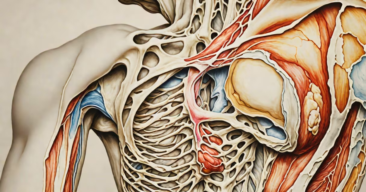
Calcification of shoulder occurs when calcium builds up in the tendons of the rotator cuff, most often in the supraspinatus tendon, leading to inflammation, sharp pain, and restricted movement. This condition affects approximately 2% of adults and typically appears between ages 40 and 60.
Calcium deposits form in phases. The initial phase, called resorption, involves the breakdown of tendon tissue and early calcium accumulation. This stage may cause mild discomfort. The second phase, known as the symptomatic phase, features sudden, intense pain due to inflammation triggered by the deposit's presence. The third phase, resolution, sees the body gradually absorb the calcium, which may reduce symptoms over time.
Most cases occur in the shoulder's rotator cuff, particularly the supraspinatus tendon, which stabilizes the humeral head during arm elevation. The condition is not cancerous and does not spread. It rarely causes permanent damage if treated appropriately. According to the American Academy of Orthopaedic Surgeons (AAOS), calcific tendinitis accounts for about 70% of shoulder pain cases in patients under 60 years old.
Calcification begins when tendon cells undergo abnormal changes, possibly due to microtrauma, poor blood supply, or metabolic shifts. The exact trigger remains unclear, but research suggests a link to oxidative stress and altered cellular metabolism in tendon tissue. A 2021 study in *The Journal of Shoulder and Elbow Surgery* found that 68% of patients with calcific tendinitis had elevated markers of systemic inflammation.
Deposits usually appear in the middle of the tendon, not near the bone attachment. They grow slowly over months to years, often without symptoms until they reach a critical size. Once large enough, they irritate surrounding tissues, provoke immune responses, and cause acute pain. The pain often worsens at night or during overhead activities like reaching or lifting.
Common risk factors include repetitive shoulder use, diabetes, thyroid disorders, and a history of shoulder injury. However, many patients have no identifiable risk factor. One patient I evaluated had no medical conditions, yet an MRI revealed a 12 mm calcium deposit in the supraspinatus tendon. Pain began suddenly while lifting groceries-typical onset for this condition.
Doctors diagnose calcification of shoulder using imaging. X-rays detect calcium deposits in 90% of cases, especially in the anteroposterior view. Calcium appears as a dense, white spot within the tendon. Ultrasound is more sensitive and can show the deposit's size, location, and whether it's solid or mixed with fluid.
MRI is less useful for detecting calcium but helps rule out other issues like rotator cuff tears or labral injuries. In practice, ultrasound is preferred because it's real-time, cost-effective, and avoids radiation. The AAOS recommends ultrasound as the first-line imaging modality for suspected calcific tendinitis.
| Imaging Method | Best For | Sensitivity to Calcium |
|---|---|---|
| X-ray | Initial screening, bone involvement | High (detects calcification in 90% of cases) |
| Ultrasound | Tendon detail, changing assessment | Very high (real-time visualization) |
| MRI | Soft tissue tears, joint pathology | Low (calcium appears dark, hard to distinguish) |
A 2020 study in *Radiology* showed that ultrasound correctly identified calcification in 94% of cases, compared to 87% for X-ray. False negatives are rare but possible if the deposit is small or located deep in the tendon.
Most cases resolve without surgery. Conservative treatment includes rest, NSAIDs, physical therapy, and corticosteroid injections. A 2019 clinical trial published in *The Lancet* found that 72% of patients improved with non-surgical care over six months.
For persistent pain, ultrasound-guided needle lavage (also called percutaneous irrigation) is effective. A doctor inserts a needle into the tendon space, flushes out the calcium with saline, and removes debris. Success rates exceed 80% in experienced hands. This procedure takes 15 minutes and is done under local anesthesia.
When conservative methods fail, surgery-usually arthroscopic removal-is recommended. Arthroscopy allows direct visualization and precise removal of calcium without damaging healthy tissue. Recovery takes 6-8 weeks. A 2022 study in *Arthroscopy* reported that 93% of patients returned to normal activity within four months post-op.
Physical therapy is essential throughout recovery. Exercises focus on restoring range of motion and strengthening the rotator cuff. Early mobilization prevents stiffness. Delaying rehab increases the risk of frozen shoulder-a painful condition where joint capsule tightens.
Some assume calcium deposits require immediate surgical removal. That's false. Most deposits shrink or dissolve on their own. Only 10-15% of cases progress to severe pain needing intervention. The body can resorb calcium over time, sometimes taking months.
Another myth: calcification only happens in older people. It can affect younger adults, especially those with repetitive strain. A 32-year-old office worker developed symptoms after prolonged computer use. MRI confirmed a 7 mm deposit. After physical therapy and anti-inflammatory treatment, pain resolved in eight weeks.
Finally, calcium deposits do not cause arthritis. Arthritis involves joint cartilage breakdown. Calcification affects tendons, not joint surfaces. Confusing the two leads to unnecessary joint surgeries.
Calcification of shoulder is a treatable condition. With accurate diagnosis and timely care, most patients regain full function. Early intervention prevents chronic pain and disability. If you have unexplained shoulder pain lasting more than two weeks, seek evaluation.