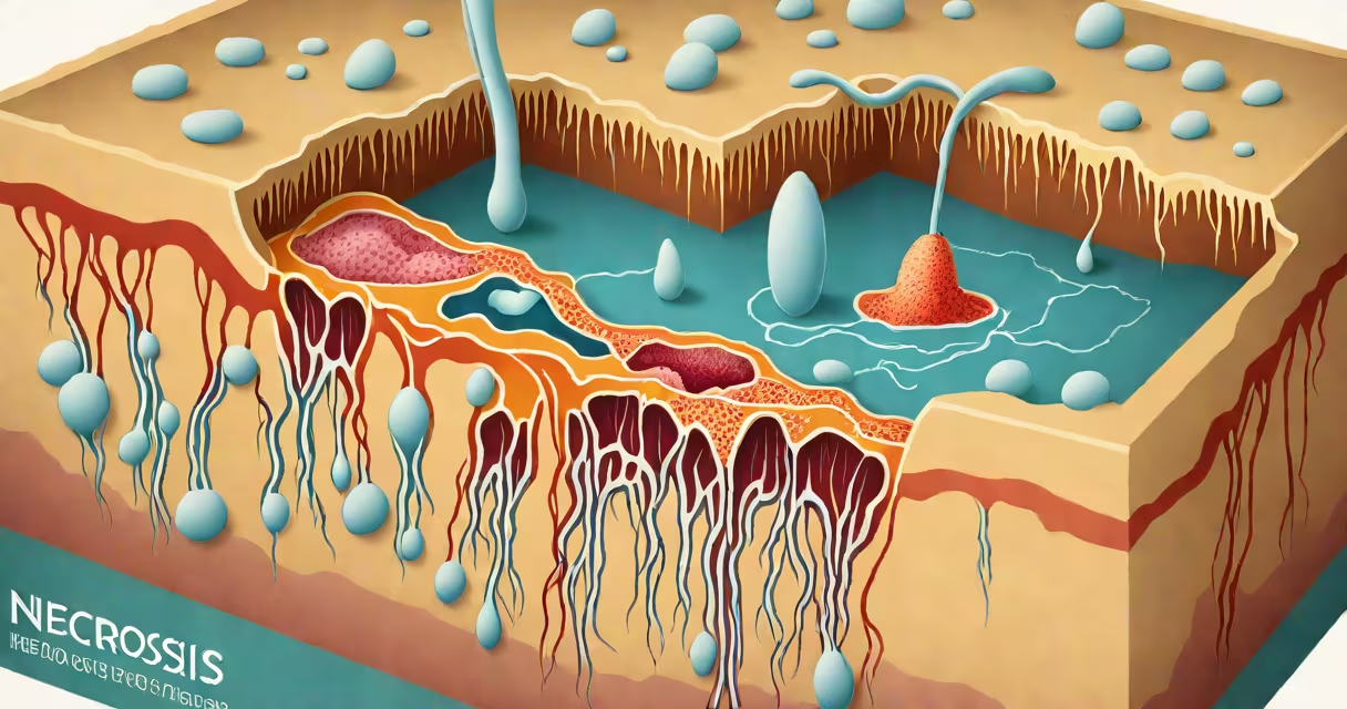
Necrosis is the pathological death of cells or tissues resulting from injury, infection, ischemia, or trauma. It occurs when cells lose their structural integrity and die through unregulated processes, unlike programmed cell death (apoptosis).
Necrosis is not a single condition but a pattern of tissue damage visible under a microscope. It typically results from external stressors such as poor blood supply, severe burns, toxins, or bacterial invasion. The affected area appears discolored-dark gray or black-and lacks cellular organization.
Medical professionals identify necrosis during biopsies, imaging scans, or surgical procedures. It signals underlying disease progression and often requires immediate intervention to prevent systemic spread. For example, a patient with a deep leg wound showing blackened tissue may have necrotizing fasciitis, a life-threatening infection causing rapid tissue death.
Necrosis begins when cells are deprived of oxygen and nutrients. This usually stems from blocked blood vessels (ischemia), which can occur during a heart attack or stroke. Without adequate perfusion, cells cannot maintain ion balance or produce energy, leading to rupture of cell membranes and leakage of intracellular contents.
The release of cellular debris triggers inflammation. Immune cells rush to the site, attempting to clear dead material. However, this response can cause collateral damage to surrounding healthy tissue. In some cases, necrotic tissue becomes a breeding ground for bacteria, increasing risk of sepsis.
A 2021 study published in *The Journal of Pathology* found that necrotic regions in tumors correlate with reduced survival rates in colorectal cancer patients. Tumor necrosis indicates aggressive growth and resistance to therapy, making it a key prognostic marker.
Pathologists classify necrosis based on appearance and underlying cause. Each type reflects different mechanisms of cell death and clinical implications.
| Type | Appearance | Common Causes | Associated Conditions |
|---|---|---|---|
| Coagulative | Gray-white, firm tissue; preserved architecture | Ischemia (e.g., heart attack) | Myocardial infarction, renal infarcts |
| Liquefactive | Soft, fluid-filled cavities | Enzymatic digestion; bacterial infection | Brain abscesses, cerebral infarcts |
| Caseous | Cheese-like, granular debris | Tuberculosis, chronic infections | Pulmonary TB, lymph node involvement |
| Fibrinoid | Amorphous, eosinophilic deposits | Immune complex deposition | Systemic lupus erythematosus, vasculitis |
| Autonecrosis | Self-dissolution due to enzyme release | Acute pancreatitis | Severe abdominal pain, elevated amylase levels |
Coagulative necrosis is most common in solid organs like the heart and kidneys. Its sign is preservation of tissue structure despite cell death-visible as a well-demarcated zone of dead tissue surrounded by inflamed borders.
Liquefactive necrosis occurs primarily in the brain. Neurons are rich in enzymes that digest proteins rapidly. After a stroke, the affected brain region liquefies into a liquid mass, forming a cyst-like cavity. This contrasts sharply with coagulative necrosis, where tissue remains structurally intact.
Caseous necrosis is pathognomonic for tuberculosis. The tissue resembles crumbly cheese under the microscope. It results from granulomatous inflammation driven by *Mycobacterium tuberculosis*. A chest X-ray showing a cavitary lesion in the upper lobe raises suspicion for active TB.
Necrosis is often confused with apoptosis-the body's normal, controlled form of cell death. Apoptosis removes aged or damaged cells without triggering inflammation. Necrosis, however, causes immune activation and tissue swelling.
Another misconception is that necrosis always indicates infection. While bacterial infections can cause necrosis, many non-infectious causes exist. For instance, radiation therapy can induce necrosis in treated tissues. A patient receiving pelvic radiotherapy for cervical cancer may develop rectal necrosis years later, even without infection.
Necrosis is irreversible. Once cells die, they do not regenerate. Surgical removal or debridement is required to prevent further complications. A 2019 review in *Nature Reviews Disease Primers* emphasized that early detection of necrosis improves outcomes in ischemic conditions. Delayed treatment increases the risk of organ failure.
A 62-year-old man with type 2 diabetes presents with a foot ulcer that won't heal. He reports numbness and tingling. On examination, the wound base is black and foul-smelling. A biopsy reveals necrotic tissue with absent blood vessels and infiltrating neutrophils.
This is wet gangrene-a form of necrosis caused by both vascular insufficiency and infection. His blood glucose levels were consistently above 200 mg/dL for months, damaging small blood vessels and impairing healing. Without prompt surgical debridement and antibiotics, the infection could spread to the bone (osteomyelitis) or enter the bloodstream.
After surgery, his wound improved. But the presence of necrosis meant he needed long-term wound care and strict glycemic control. Monitoring for signs of recurrence became essential.
Necrosis is a red flag in pathology reports. It suggests a serious underlying issue requiring urgent attention. Radiologists look for areas of low density on CT scans or signal changes on MRI that indicate necrotic tissue.
In oncology, tumor necrosis is a sign of poor prognosis. The American Joint Committee on Cancer (AJCC) includes necrosis as a staging criterion for certain cancers, including soft tissue sarcomas. Presence of necrosis increases stage classification and influences treatment decisions.
For clinicians, recognizing necrosis early can prevent limb loss, reduce hospital stays, and improve survival. A 2020 study in *The New England Journal of Medicine* showed that patients with acute limb ischemia who received revascularization within six hours had lower amputation rates than those treated later.
When evaluating a patient, doctors consider history, physical exam, lab tests like lactate and CRP, and imaging. Elevated lactate suggests poor tissue perfusion. High CRP indicates ongoing inflammation. Together, these markers help confirm whether necrosis is present.
Understanding what necrosis means in medical terms enables accurate diagnosis and timely intervention. It is not a standalone diagnosis but a sign of deeper dysfunction-be it vascular, infectious, or neoplastic.
Necrosis is a critical concept in medicine. It defines tissue death beyond repair and demands clinical action. Recognizing its patterns, causes, and consequences allows for better patient outcomes across specialties-from cardiology to dermatology to oncology.