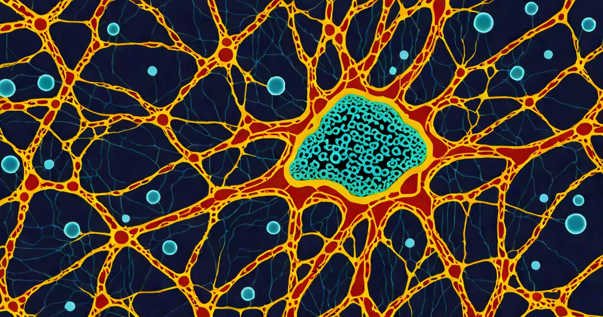
Vimentin is a type of intermediate filament protein that forms a flexible, resilient network within the cytoplasm of mesenchymal cells, providing mechanical stability and organizing cellular structures. It is a key component of the cytoskeleton and is expressed in cells derived from the mesoderm, including fibroblasts, endothelial cells, and certain immune cells.
Intermediate filaments like vimentin are distinct from actin microfilaments and microtubules. They do not participate in cell motility or intracellular transport but instead serve as internal scaffolds. Vimentin filaments are 10-12 nm in diameter and assemble into stable, rope-like structures that resist mechanical stress. These filaments connect to desmosomes and focal adhesions, anchoring the nucleus and organelles in place during cell movement and division.
Human vimentin is encoded by the *VIM* gene on chromosome 21. The protein consists of a central rod domain flanked by non-helical head and tail domains. This modular design allows for changing assembly and disassembly in response to cellular signals. Phosphorylation of specific serine residues during mitosis causes vimentin to depolymerize, enabling proper spindle orientation and daughter cell separation.
Vimentin maintains cell integrity under physical strain. In fibroblasts, which are constantly exposed to stretching forces in connective tissues, vimentin networks absorb shear stress and prevent membrane rupture. Without vimentin, cells show increased fragility and impaired wound healing in animal models.
The protein also plays a role in organelle positioning. Vimentin filaments extend from the perinuclear region toward the cell periphery, forming tracks along which mitochondria and lysosomes move. Disruption of vimentin leads to mislocalization of these organelles, affecting energy distribution and waste clearance.
A 2021 study using cryo-electron microscopy revealed that vimentin filaments adopt a staggered helical arrangement, with each monomer contributing to a continuous fiber. This structure allows for flexibility without breaking, unlike rigid microtubules or contractile actin bundles.
Vimentin belongs to the type III intermediate filament family. Other members include desmin (muscle cells), GFAP (astrocytes), and peripherin (neurons). Each type is expressed in specific cell lineages and serves tissue-specific roles.
| Cell Type | Primary Intermediate Filament | Function |
|---|---|---|
| Fibroblast | Vimentin | Mechanical resilience, wound repair |
| Smooth muscle | Desmin | Myofiber integrity, contraction coordination |
| Neuron | Neurofilament (type IV) | Diameter regulation, axonal transport |
| Brain astrocyte | GFAP | Glial scar formation, metabolic support |
Vimentin expression is normally absent in epithelial cells, which rely on keratins. However, during epithelial-mesenchymal transition (EMT)-a process critical in embryonic development and cancer metastasis-epithelial cells begin expressing vimentin. This shift marks a loss of polarity and gain of migratory capacity.
Vimentin is widely used as a biomarker in immunohistochemistry. Its presence in tumor biopsies indicates mesenchymal origin or EMT activation. For example, in breast cancer, vimentin-positive cells correlate with higher grade, resistance to chemotherapy, and increased risk of recurrence.
A 2019 study published in *Nature Cell Biology* showed that vimentin levels rise in activated hepatic stellate cells during liver fibrosis. Researchers used vimentin staining to track disease progression in mouse models and identified it as a potential therapeutic target.
In clinical practice, vimentin is often tested alongside cytokeratins. A biopsy positive for vimentin and negative for cytokeratin suggests a sarcoma rather than a carcinoma. This distinction guides treatment decisions, as sarcomas require different chemotherapy regimens.
Vimentin is not involved in cell division mechanics. Unlike tubulin, which forms the mitotic spindle, vimentin disassembles during mitosis but does not contribute to chromosome segregation. Confusing it with microtubule proteins leads to errors in interpreting cell cycle dynamics.
Another misconception is that vimentin expression always indicates malignancy. While elevated vimentin occurs in many aggressive tumors, it is also normal in healing wounds and inflamed tissues. A single positive stain does not confirm cancer-context matters.
Vimentin is not a universal marker of stemness. Some mesenchymal stem cells express vimentin, but so do differentiated fibroblasts. The protein's presence alone cannot distinguish between progenitor and mature cell states.
A 52-year-old patient presents with a soft-tissue mass in the thigh. Imaging shows a 6 cm lesion with irregular borders. A core biopsy is taken and stained with hematoxylin and eosin (H&E), revealing spindle-shaped cells with high nuclear-to-cytoplasmic ratios.
Immunohistochemistry panels are run. The tumor cells are negative for cytokeratin (AE1/AE3), ruling out epithelial origin. They are strongly positive for vimentin. CD34 is negative, excluding gastrointestinal stromal tumor (GIST). S100 is negative, eliminating melanoma.
Based on this profile-vimentin-positive, cytokeratin-negative-the diagnosis is malignant fibrous histiocytoma (now classified as undifferentiated pleomorphic sarcoma). The patient undergoes wide local excision and adjuvant radiation. Follow-up imaging at six months shows no recurrence.
This case illustrates how vimentin serves as a diagnostic anchor in differential pathology. Without it, distinguishing sarcoma from carcinomas would rely on less specific markers.
I observed vimentin staining patterns in a dermatopathology lab during a rotation. A skin biopsy from a patient with chronic inflammation showed intense vimentin expression in dermal fibroblasts surrounding granulomatous lesions. The pattern mirrored the inflammatory zone, confirming active tissue remodeling. This visual correlation helped me understand how vimentin reflects functional state, not just identity.
Vimentin is a structural protein essential for maintaining cell shape and resisting mechanical stress in mesenchymal cells. It functions as a scaffold within the cytoskeleton and serves as a critical biomarker in pathology. Its expression in cancer and fibrotic diseases makes it valuable for diagnosis and research. Understanding what is vimentin clarifies its role beyond a simple label-it is a changing player in cell architecture and disease progression.