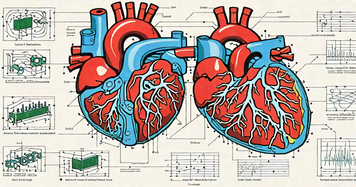
Fluid in the heart, medically known as pericardial effusion, occurs when excess fluid builds up in the pericardial space-the area between the heart's outer lining (pericardium) and the heart muscle itself. This fluid can impair heart function if it compresses the organ, leading to life-threatening complications.
Pericardial effusion is not a disease itself but a sign of an underlying issue. It can result from infection, inflammation, trauma, cancer, or heart failure. The fluid acts as a cushion under normal conditions, but when excessive, it disrupts the heart's ability to fill properly during each beat.
The condition affects approximately 1 in 1,000 adults seen in emergency departments, according to the American Heart Association. It is often detected incidentally on imaging scans or during evaluation of unexplained fatigue, chest pain, or breathing difficulties.
The pericardium is a double-layered sac surrounding the heart. Under normal conditions, a small amount of lubricating fluid exists between the layers to reduce friction during heartbeats. When this balance is disrupted, fluid accumulates due to increased production or decreased absorption.
Common causes include viral infections (such as coxsackievirus), autoimmune disorders like lupus, kidney failure requiring dialysis, and complications from heart surgery. Less frequently, malignancies such as lymphoma or metastatic breast cancer can invade the pericardium and trigger fluid leakage.
A 2021 study published in *Circulation* found that 60% of patients with significant effusions had identifiable causes, with heart failure being the most common diagnosis among hospitalized individuals.
Effusions are categorized by volume and clinical impact:
| Type | Volume | Clinical Impact | Common Causes |
|---|---|---|---|
| Small | <100 mL | Often asymptomatic; detected via echocardiogram | Viral illness, post-surgical changes |
| Moderate | 100-500 mL | Possible symptoms: mild shortness of breath, chest tightness | Inflammation, hypothyroidism |
| Large | >500 mL | Significant risk of cardiac tamponade | Trauma, cancer, severe infections |
Cardiac tamponade-a medical emergency-occurs when fluid pressure restricts heart filling. It presents with pulsus paradoxus (exaggerated drop in blood pressure during inhalation), low blood pressure, and muffled heart sounds. Without treatment, it can lead to shock and death within hours.
One non-obvious fact: not all fluid around the heart is pathological. Up to 15% of healthy individuals show trace effusions on routine echocardiograms, especially older adults or those with chronic kidney disease. These findings typically don't require intervention unless symptoms develop.
Doctors diagnose pericardial effusion using echocardiography, which provides real-time images of the heart and measures fluid volume. CT scans and MRI offer additional detail in complex cases.
Many people confuse fluid in the heart with pulmonary edema-fluid in the lungs-which causes similar symptoms like breathlessness and coughing. However, pulmonary edema stems from left-sided heart failure, while pericardial effusion affects the heart's external environment.
Another misconception is that fluid in the heart always requires drainage. In reality, small effusions may resolve on their own. A 2019 review in *The New England Journal of Medicine* noted that 40% of patients with mild effusions experienced spontaneous resolution over six months without treatment.
A 62-year-old man presented to the ER with progressive shortness of breath and fatigue. He had no history of heart disease but reported a recent upper respiratory infection. On physical exam, his blood pressure dropped from 130/80 mmHg to 90/60 mmHg during inspiration-a sign of pulsus paradoxus.
An echocardiogram revealed a 450 mL effusion with signs of impaired ventricular filling. Blood tests showed elevated inflammatory markers. After ruling out malignancy and infection, doctors diagnosed idiopathic pericarditis with secondary effusion.
He was treated with NSAIDs and colchicine. Over two weeks, the fluid resolved. Follow-up imaging confirmed near-complete clearance. His symptoms improved fully within four weeks.
Symptoms such as sudden chest pain, dizziness, rapid breathing, or swelling in the legs may signal worsening effusion. Immediate care is needed if confusion, cold extremities, or fainting occur-signs of impending shock.
Fluid in the heart is not a standalone diagnosis. It reflects an underlying process affecting the pericardium. Most cases are treatable when caught early. Not all effusions require intervention-small ones may disappear without action.
Accurate diagnosis depends on imaging and clinical context. Do not assume fluid means cancer or immediate danger. But do not ignore symptoms either. Early detection prevents complications like tamponade.
Understanding what fluid in the heart means helps distinguish between harmless findings and serious conditions. Always consult a cardiologist if imaging shows effusion, especially with symptoms.