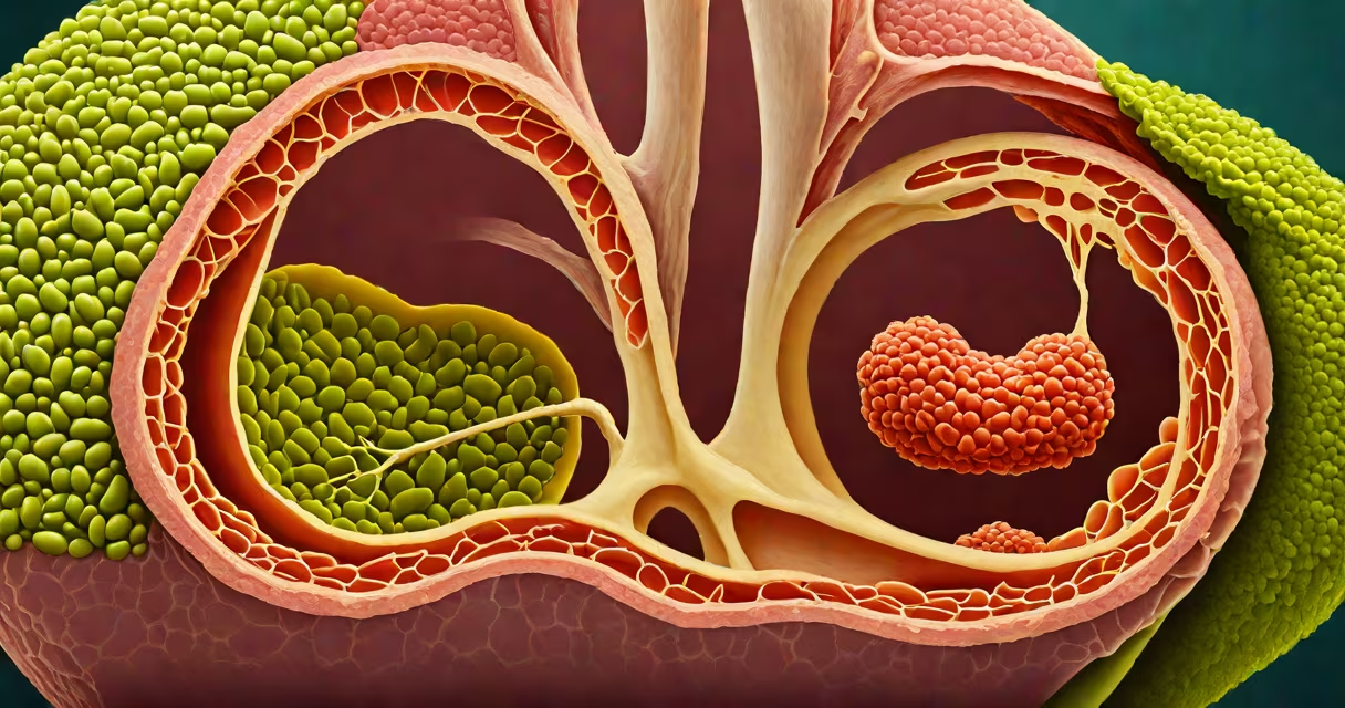
The human gallbladder is a small, pear-shaped organ situated beneath the liver in the right upper quadrant of the abdomen, tucked into a shallow fossa on the inferior surface of the liver's right lobe. It lies just below the diaphragm and is anchored by connective tissue to the abdominal wall.
Its position is consistent across adults: approximately 10 cm below the right costal margin and 5 cm lateral to the midline. This anatomical placement makes it vulnerable to trauma during blunt abdominal injury and explains why pain from gallbladder disease often presents in the right upper quadrant, sometimes radiating to the right shoulder due to referred pain via the phrenic nerve.
Despite its small size-typically 7-10 cm long and 3-4 cm wide-the gallbladder plays a critical role in fat digestion. It concentrates and stores bile produced by the liver, releasing it into the duodenum through the common bile duct during meals to emulsify dietary fats.
Bile flows from the liver through the right and left hepatic ducts, which merge to form the common hepatic duct. This duct joins the cystic duct from the gallbladder to create the common bile duct, which empties into the duodenum at the ampulla of Vater. The gallbladder itself does not produce bile-it only stores and concentrates it, reducing water content by up to 90% during storage.
When fatty food enters the duodenum, the hormone cholecystokinin (CCK) triggers gallbladder contraction. This forces concentrated bile into the duodenum, where it acts on triglycerides to increase their surface area for enzymatic breakdown by pancreatic lipase. Without this process, fat absorption drops, leading to steatorrhea-fatty, foul-smelling stools.
A study published in *Gastroenterology* found that 60-70% of patients with symptomatic gallstones report pain in the right upper quadrant, confirming the clinical relevance of the organ's precise location. Pain may worsen after high-fat meals, a sign of biliary colic.
While most people have a single gallbladder, rare anatomical variants exist. Some individuals have accessory gallbladders (up to 0.3% incidence), which are usually asymptomatic and discovered incidentally during imaging or surgery. Others may have a double gallbladder or a gallbladder that sits higher in the abdomen, potentially complicating laparoscopic removal.
During laparoscopic cholecystectomy-the standard treatment for symptomatic gallstones-surgeons identify the gallbladder by locating the cystic duct and artery. The Calot's triangle, formed by the cystic duct, common hepatic duct, and the cystic artery, serves as a key landmark. Misidentification of structures in this region leads to complications such as bile duct injury, which occurs in 0.3-0.5% of cases, according to the American Society for Gastrointestinal Endoscopic Surgeons (ASGE).
Post-surgical patients often report changes in digestion, particularly after consuming large amounts of fat. A 2021 study in *The Journal of Clinical Gastroenterology* noted that 15% of patients experienced increased diarrhea or bloating after cholecystectomy, though symptoms typically resolve within six months.
Many assume the gallbladder is essential for survival. This is false. Humans can live normally without a gallbladder. After surgical removal, bile flows continuously from the liver into the small intestine rather than being stored and released in bursts. The body adapts over time, though some individuals develop postcholecystectomy syndrome, characterized by persistent abdominal pain, nausea, or diarrhea.
Another misconception is that gallbladder pain always indicates gallstones. While stones are the most common cause of biliary colic, conditions like acalculous gallbladder disease-where the gallbladder fails to contract properly despite no stones-can mimic the same symptoms. This condition affects 10-15% of patients undergoing cholecystectomy who have normal ultrasound results.
The gallbladder is frequently confused with the pancreas or spleen. The pancreas lies deeper in the upper abdomen, behind the stomach, and produces insulin and digestive enzymes. The spleen is located in the left upper quadrant and filters blood. Imaging studies such as ultrasound or CT scans are required to distinguish these organs accurately.
| Organ | Location | Primary Function |
|---|---|---|
| Gallbladder | Right upper quadrant, under liver | Stores and concentrates bile |
| Pancreas | Deep upper abdomen, behind stomach | Produces insulin and digestive enzymes |
| Spleen | Left upper quadrant, near stomach | Filters blood and supports immune function |
During a routine physical exam, the gallbladder cannot be palpated unless enlarged or inflamed. A positive Murphy's sign-pain upon deep inspiration during palpation of the right upper quadrant-is a clinical indicator of acute cholecystitis. This test has a sensitivity of 70% and specificity of 85% for detecting inflammation, according to guidelines from the American College of Radiology.
I once observed a patient with severe right upper quadrant pain who had normal lab values and a negative ultrasound. The diagnosis was later confirmed via HIDA scan, which showed no gallbladder ejection fraction-indicating functional failure without structural abnormalities. This case underscored that location alone doesn't confirm pathology; function matters too.
Knowing the exact location of the gallbladder helps explain why symptoms like pain, nausea, and jaundice arise in specific regions of the abdomen. It also clarifies why diagnostic tools like ultrasound and HIDA scans rely on spatial relationships between the liver, gallbladder, and bile ducts.